UNT Ion Beam Laboratory (IBL)
UNT Ion Beam Laboratory (IBL)
Dr. Bibhu Rout (bibhu@unt.edu)
Dr. Duncan Weathers (weathers@unt.edu)
Mission
The Ion Beam Laboratory (IBL) utilizes medium energy (10 keV-12 MeV) ion accelerators for multi-disciplinary research and development utilizing a wide range of analytical and modification research techniques in basic and applied physics. IBL students develop skills that can be attractive to academia and industry in areas such as biophysics, semiconductors, solar cells, nuclear energy, environmental science, and other areas of materials science. IBL is also a recognized leader in high energy focused ion beam (HEFIB) technology development and applications.
History
In 1986, the accelerator group at the University of North Texas received a Texas Advanced Technology Research Grant to acquire a National Electrostatics Corporation Model 9-SDH 3 million-volt tandem Pelletron®-type accelerator which was installed in December 1987. UNT also provided funds to construct an addition to the Ion Beam Modification and Analysis Laboratory (IBMAL) to house the tandem accelerator.
The 200 kV Cockcroft-Walton accelerator and one of the 2.5 MV Van de Graaff's was located in a 78 square meter laboratory while the other three accelerators were located in a 450 square meter adjacent laboratory. The Cockcroft-Walton accelerator was used for surface analysis measurements of materials by sputter-initiated resonance ionization spectroscopy (SIRIS). The 3 MV tandem accelerator, which is still one of the workhorses of the laboratory, was purchased with an rf charge exchange ion source (alphatross) and a Cs sputter-type ion source (SNICS).
A third, ultra-clean, microbeam, raster-scanning, depth-profiling, Cs-sputtering ion source was constructed for the 3 MV tandem accelerator for Trace Element Accelerator Mass Spectroscopy (TEAMS). TEAMS was used to determine impurities in materials at parts per billion (ppb) by depth profiling techniques. In 2021, the TEAMS system was decommissioned to make room for new research instrumentation.
In 2011, a 3 MV single-ended Pelletron® accelerator was moved from the UNT Material Science department to the Physics department ion beam laboratory. This accelerator system was upgraded with a new state-of-the-art digital control system and high stability terminal control. Subsequent improvements to the laboratory included design, fabrication and installation of a high energy focused ion microprobe on one of the beamlines on the 3 MV single-ended Pelletron. More recently, IBL has developed a patented system to allow detection and quantification of trace amounts of low-mass elements (mass>boron) in organic and inorganic matrices.
In 2022 the name of the laboratory was changed to the UNT Ion Beam Laboratory (IBL) to encompass more completely the wide range of ongoing research. Current instrumentation at IBL includes two National Electrostatics Corporation accelerators; a 3 MV 9SH single-ended Pelletron® and a 3 MV SDH2 tandem Pelletron® accelerator. The 200 kV Cockcroft Walton and 2.5 MV High Voltage Engineering single-ended accelerators are situated in a separate laboratory at IBL. IBL yearly maintenance and operating expenses, students and postdocs are supported by research grants and service work. Typical IBL personnel includes three full time faculty (Gary Glass (Director), Dr. Bibhudutta (Associate Director) and Dr. Duncan Weathers along with six graduate student research assistants. Figure 1 illustrates the layout of the accelerator area at IBL while Figure 2 shows a view of all the beamlines.
The industrial interactions with the IBL, which were begun in the late 1970's, have continued over the past few years. One of the reasons for the close interactions with industry has been the International Conference on the Application of Accelerators in Research and Industry (CAARI), which is co-sponsored by UNT. This conference has been held since 1976, in even numbered years, missing only 2020 due to the COVID-19 worldwide pandemic. The 27th CAARI will be held on 21-26 July 2024. https://caari-sneap.com in Denton, TX. The 2024 Symposium of Northeast Accelerator Personnel (SNEAP) will be jointly held with CAARI.
IBL research programs have been supported by a number of agencies, including the National Science Foundation, the National Institutes of Health, the Office of Naval Research, the Robert A. Welch Foundation, the University of North Texas, the State of Texas Advanced Research Programs, and a number of industrial sponsors.
 Accelerator Area")
Figure 1. Ion Beam Laboratory (IBL) Accelerator Area

Figure 2. View of beamlines at IBL
Research Applications of Ion Beams
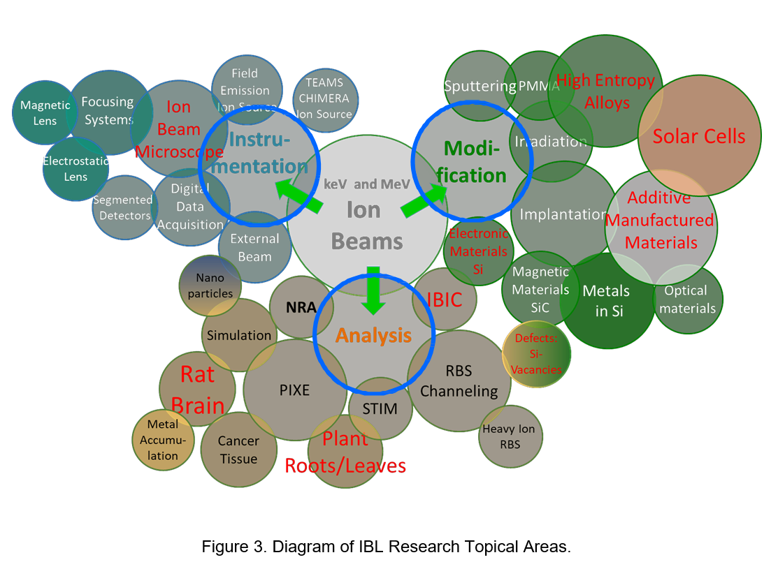
IBL currently has ongoing research in a variety of topics. These topics can be categorized in one of three general areas (see Figure 3): 1) Analysis, 2) Modification and 3) Instrumentation. Some of the more biophysics, focused ion beam systems development, biophysics, environmental science, laboratory astrophysics, and materials. Capabilities include a number of analytical ion beam techniques including Particle Induced X-ray Emission Spectroscopy (PIXE), Rutherford Backscattering Spectroscopy (RBS), Nuclear Reaction Analysis (NRA), Hydrogen Forward Scattering (HFS), Ion Channeling (IC) and ion implantation. Some of the microprobe techniques available at IBL include Ion Beam Induced Luminescence (IBIL), Ion Beam Induced Charge Collection (IBICC), microbeam PIXE and RBS, and ion microlithography.
1) Ion Beam Analysis
Particle Induced X-ray Emission Spectroscopy (PIXE)
PIXE has proven to be a reliable and sensitive technique for the detection and quantification of trace elements with atomic numbers in the range of 13-92. PIXE relies upon the relatively high probability that an energetic (1-3 MeV) ion (usually a proton) can undergo an inelastic collision with the electrons in an atom that results in the ionization of an inner-shell electron. The resulting vacancy will be filled by other electrons cascading from higher shells, releasing energy that can be emitted as one or more x-rays characteristic of the atom from which it originates. Detecting and quantifying these characteristic X-rays can provide information to determine elemental constituents and the quantity of those constituents in a sample with the identified peaks in the spectrum originating from X-rays of specific shells within the atom. The number of X-rays, defined as the yield, detected in a peak corresponding to a particular shell from a specific atom is proportional to the concentration of that atom in the sample. With the recent development of ultra-thin entrance windows for detectors, the range of measurable elements has been extended to atomic numbers as low as boron, however damage to the detector from backscattered incident ions can significantly reduce the useful life of the detector. An absorber of appropriate thickness can be placed in front of the detector to eliminate most of these backscattered ions. However, the use of an absorber will also preclude the measurement of low energy X-rays emitted by the low-Z elements. Figure 4 shows a typical PIXE spectrum obtained from a sample of dust captured by falling raindrops.
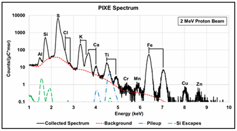
Figure 4. Typical PIXE spectrum from dust absorbed in rain. The secondary electron and proton bremsstrahlung background radiation dominates the low energy portion of the spectrum. The dotted red line indicates the calculated bremsstrahlung background. An absorber placed between the target and the X-ray detector can reduce this component.
Proton Microscopy
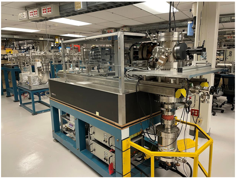
Magnetic quadrupole lenses arranged as doublets, triplets, quadruplets, quintuplets, sextuplets etc. can be used to focus 1-3 MeV ion beams (primarily protons) to µm-sized (or less) beam spots (microbeam). The arrangement of the ion source, accelerator, slits, lenses and endstation constitute a complete “focused ion beam” system. In order to achieve a small and bright beam focus, the lenses must be fabricated with high precision having both mechanical and magnetic symmetry if possible. The focused beam is scanned across a sample using magnetic or electrostatic deflection systems with one of two objectives: (1) Mapping recorded primary or secondary “events” as a function of beam position to provide “images” of the surface or structure, or (2) controlled irradiation of a sample to produce micro-patterns. The UNT Scanning Proton Microprobe (SPM) system is shown in Figure 5.
UNT Magnetic Quadrupole Quadruplet Microprobe
Figure 5. The unique IBL high energy focused ion beam (HEFIB) system at the University of North Texas can focus 1–3 MeV protons to spot sizes less than 1×1μm2 as well as 1–4.5 MeV carbon and silicon ion beams to spot sizes of 1.5×1.5μm2. This allows very high resolution 2-dimensional mapping and quantification of multiple elements simultaneously. The system is comprised of 2 sets of magnetic quadrupole doublets. The system can be operated in several different modes: doublet, triplet and quadruplet.
Microbeam PIXE
The Proton Induced X-ray Emission (PIXE) microprobe technique is illustrated in Figure 6(a). Samples are scanned by a rastered proton beam in vacuo by characteristic X-ray emissions. These emissions are unique for individual elemental constituents and can be used to quantify the concentration of the individual elements. Figures 6 (b) and (c) demonstrate the ability of the microPIXE technique to provide cellular-level profiles of several elements of interest. The analysis software allows the user to re-analyze any area of interest with regard to element(s) of interest and/or orientation of line scans without the need to again expose the sample to the microbeam. Additionally, correlation techniques such as scatter diagrams using intensity relations (ratios) between the various elemental maps, can lead to the calculation of chemical phase maps. Figure 7 illustrates this correlation technique for Fe and Si in an environmental dust sample. This is one of the major innovative techniques available at IBL. All these analytical measurements were performed non-destructively on systems with minimal sample preparation. These correlation maps can be performed for in-situ biological samples to provide in-chemical state (phase) maps.
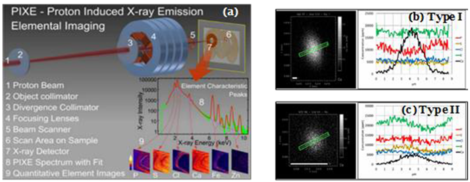
Figure 6. (a) PIXE quantitative elemental mapping principles. A high energy proton beam (1) focused (4) to 1×1 μm² is scanned over sample areas of interest (6). Characteristic X-ray emissions from proton excitation of sample constituents recorded by the X-ray detector(7), resulting in a spectrum of X-rays (8). Specialized software determines quantitative elemental distributions (9) over individual micron-square pixels in the scanned area. Dry mass constituents are determined by RBS. (b and c). Cellular level microPIXE scans of Type I (b) and Type II (c) rat brain neurons. Line scans indicate different profiles of P, S, Cl and K for Type I and Type II neurons.
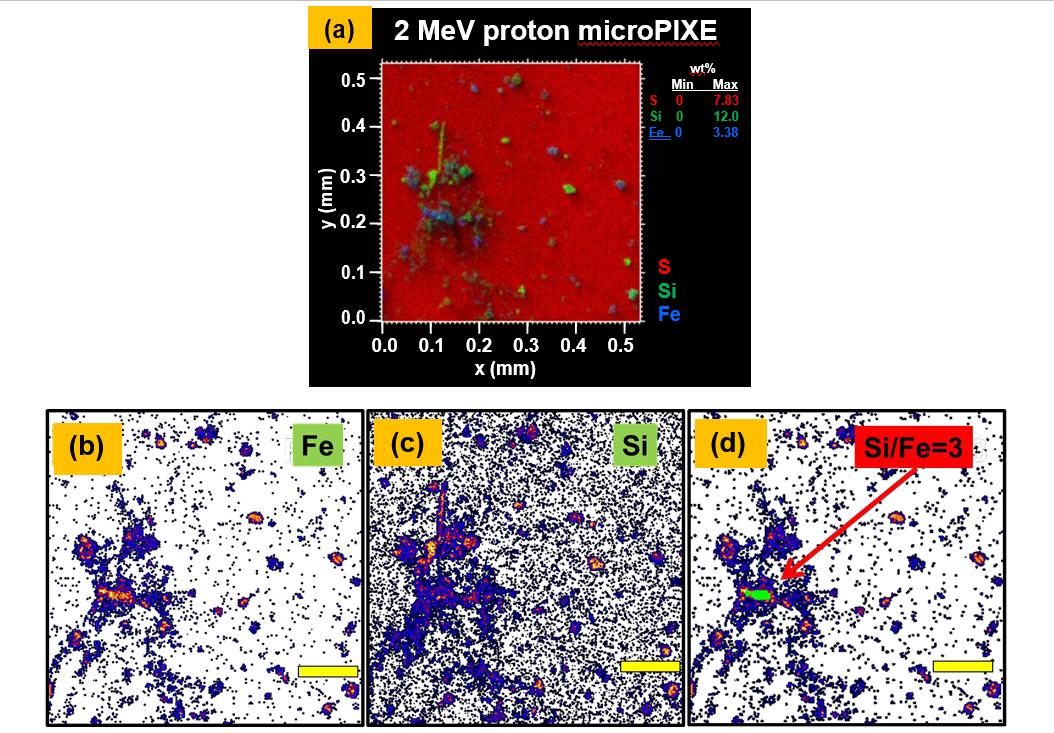
Figure 7. Demonstration of phase (correlation) mapping technique. (a) 500 mm x 500 mm microPIXE image of dust particles on polysulfone membrane, (b)-(d) are x-ray maps of iron and silicon, resp. The area in green in Figure (d) indicates region with silicon concentration is 3 × iron concentration. (Yellow scale bars = 100 µm.)